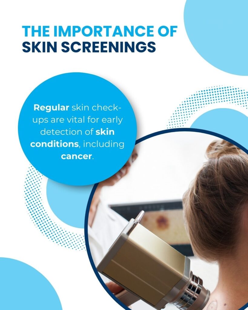
Skin cancer, particularly malignant melanoma (MM), is a growing health concern in the UK and worldwide. With over 16,000 new cases diagnosed annually in the UK alone, early and accurate detection is critical to improving patient outcomes and reducing mortality rates. However, diagnosing MM remains a challenge due to its similarity to benign lesions, leading to unnecessary biopsies, increased patient anxiety, and strain on healthcare resources.
Reflectance Confocal Microscopy (RCM) is emerging as a game-changing diagnostic tool in dermatology. This non-invasive imaging technology provides real-time, high-resolution views of skin lesions at a cellular level, allowing for more precise identification of malignant melanomas and other suspicious lesions. By integrating RCM into clinical workflows, The Skincare Network is at the forefront of revolutionising skin cancer diagnosis in the UK.
Traditional Diagnostic Methods vs. RCM
- Clinical Examination
The clinical examination is often the first step in evaluating suspicious skin lesions. Dermatologists visually inspect moles or marks on the skin, assessing their size, shape, colour, and texture. While this method is essential for initial screening, it has significant limitations:
- Sensitivity and specificity rates are relatively low compared to advanced diagnostic tools.
- Many benign lesions are misclassified as malignant, leading to unnecessary excisions.
- Dermoscopy
Dermoscopy enhances clinical examination by using magnification and polarised light to provide a detailed view of skin structures beneath the surface. It is widely regarded as a key tool for evaluating pigmented lesions.
- Strengths: Dermoscopy improves sensitivity compared to visual inspection alone and helps identify patterns indicative of melanoma.
- Weaknesses: Specificity remains limited for equivocal lesions, often resulting in over-diagnosis of potential malignancies and unnecessary biopsies.
- Reflectance Confocal Microscopy (RCM)
RCM represents a significant advancement in skin cancer diagnostics by offering real-time imaging at a cellular level without invasive procedures:
- It provides high-resolution images of the dermoepidermal junction (DEJ), where melanoma typically develops, allowing for more precise identification of malignant cells.
- Clinical trials have demonstrated that RCM achieves higher sensitivity (94.2%) and specificity (83.0%) than dermoscopy alone.
- The number needed to treat (NNT)—the average number of lesions that need to be excised to find one melanoma—is significantly reduced with RCM:
- Clinical examination: NNT = 3.86
- Dermoscopy: NNT = 2.96
- RCM: NNT = 1.30
By reducing unnecessary biopsies while maintaining high diagnostic accuracy, RCM addresses key limitations of traditional methods.
Key Findings from a Recent Studies
A recent prospective observational trial, led by The Skincare Network in collaboration with the University La Sapienza, Rome, the School of Medicine and Population Health at the University of Sheffield, and Cellular Pathology Services, Watford, assessed the effectiveness of RCM in diagnosing malignant melanoma and lentigo maligna (LM) within a UK cohort. The results, published in the British Journal of Dermatology, provide valuable insights into RCM’s performance.
Key findings from the study include:
- Sensitivity and Specificity: For diagnosing MM and LM, RCM achieved a sensitivity of 94.2% (95% CI 87.0–98.1) and a specificity of 83.0% (95% CI 79.9–85.8). In comparison, clinical examination had a sensitivity of 62.8% and specificity of 63.1%, while dermoscopy showed a sensitivity of 91.9% and specificity of 42.0%. These results confirm that RCM improves diagnostic accuracy compared to clinical examination and dermoscopy alone.
- Negative Predictive Value (NPV): RCM demonstrated a high NPV of 99.1% (95% CI 97.9–99.6), indicating that when RCM suggests a lesion is benign, there is a very low chance of it being malignant.
- Reduction in Number Needed to Treat (NNT): The study highlighted that RCM significantly reduces the NNT, which decreased from 3.86 with clinical examination to 2.96 with dermoscopy and further to 1.30 with RCM. This reduction underscores the potential of RCM to minimise unnecessary biopsies.
These findings support the use of RCM in UK pigmented lesion clinics by dermatologists trained in RCM, offering a reliable method for diagnosing MM and LM.
Benefits of RCM for Patients
RCM offers several advantages that make it an invaluable addition to modern dermatology practices:
- Non-Invasive Procedure: Unlike biopsies, RCM involves no cutting or tissue removal, making it painless and stress-free for patients.
- Immediate Results: Patients receive instant feedback on whether their lesion is benign or requires further investigation, reducing waiting times and anxiety associated with biopsy results.
- Minimises Unnecessary Excisions: By accurately identifying benign lesions, RCM spares patients from scarring and discomfort caused by avoidable surgical procedures.
- Ideal for Complex Cases: RCM is particularly effective for diagnosing challenging conditions like lentigo maligna or lesions located in cosmetically sensitive areas such as the face.
Why Choose The Skincare Network for RCM?
The Skincare Network is uniquely positioned to offer advanced diagnostic services using Reflectance Confocal Microscopy. Here’s why patients trust us for their skin health needs:
- Expertise: Our dermatologists are trained in RCM technology through international programmes, ensuring the highest level of proficiency in interpreting confocal images.
- State-of-the-Art Equipment: We utilise VivaScope systems—the same technology used in leading European centres—to deliver accurate and reliable results.
- Comprehensive Care: We integrate RCM into our clinical workflows alongside clinical examination and dermoscopy to provide a thorough evaluation of suspicious lesions.
As one of the few clinics in the UK offering RCM diagnostics, The Skincare Network is committed to delivering cutting-edge care that prioritises patient safety and comfort.
What Patients Can Expect During an RCM Evaluation
- Initial Consultation: Patients undergo a clinical examination followed by dermoscopy to assess their skin lesions comprehensively.
- RCM Imaging: Suspicious lesions are scanned using advanced confocal microscopy equipment, providing high-resolution images at a cellular level. The procedure typically takes about 10 minutes per lesion and is entirely non-invasive.
- Diagnosis: Immediate feedback is provided on whether the lesion is benign or requires further investigation. Biopsies or excisions are performed only if necessary, ensuring minimal disruption to patients’ lives.
This streamlined process allows patients to receive accurate diagnoses quickly while avoiding unnecessary procedures.
Conclusion
Reflectance Confocal Microscopy (RCM) is transforming skin cancer diagnosis by improving accuracy, reducing patient anxiety, and minimising unnecessary excisions. Its ability to provide real-time imaging at a cellular level makes it an invaluable tool for dermatologists worldwide. As demonstrated by The Skincare Network’s pioneering UK-based study, RCM delivers superior diagnostic performance compared to traditional methods like clinical examination and dermoscopy.
Experience Advanced Skin Cancer Care Today
The Skincare Network is proud to be among the few clinics in the UK offering Reflectance Confocal Microscopy (RCM) technology for skin cancer diagnosis. Our expert dermatologists are dedicated to providing personalised care using state-of-the-art equipment.
If you have concerns about suspicious moles or lesions, don’t wait—early detection saves lives. Book an appointment today to experience the benefits of RCM firsthand.
Visit www.skincarenetwork.co.uk or call us to schedule your consultation with our specialists. Take control of your skin health today!
